What is the Biceps Muscle Tendon Unit?
The biceps muscle tendon unit is anatomically located in the front of the arm. The muscle belly is comprised of two muscle bellies: a long head (LHB) and a short head thus explaining the name “biceps”. The “biceps” crosses both the shoulder and elbow joints and thus the action of the muscle has an effect on both joints. The biceps is responsible for allowing one to flex or bend the elbow upwards towards the shoulder (Fig 1). It is an important muscle providing strength to allow one to lift objects with the palm facing upwards as in performing a curl. In addition the biceps, is the most powerful muscle that allows one to rotate their palm upward when the elbow is bent at a 900 angle. An example of this activity would the tightening of a screw or turning the key in an ignition.
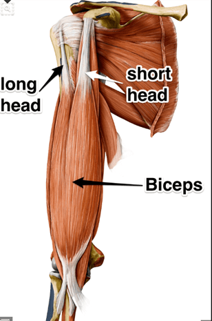
Figure 1 Anatomy of Biceps Muscle Tendon Unit
What are Common Injuries of the Biceps Tendon?
Commonly, injuries to the biceps involve either the long head located near the shoulder and distal insertion site at the elbow. The hallmark symptom is pain. Inflammation of the sheath surrounding the long head tendon is commonly referred to as biceps tendonitis in the shoulder. The tendon can be directly inspected and visualized by placing a camera called an “arthroscope” in the shoulder joint. Degeneration and thickening of the long head or the distal biceps tendon can occur over time with aging and overuse and this is referred to as biceps tendinosis. Tears, both partial and complete, can affect both the long head and the distal insertion at the elbow. Complete tears of the LHB can occur due to age related chronic degeneration and tearing (Fig 2), whereas distal biceps rupture at the elbow nearly always occur in the setting of trauma that occurs when attempting to lift a heavy object. Lastly instability of the long head of the biceps can also occur. Because the LHB runs in a bony groove between the rotator cuff muscles, injuries the ligaments maintaining the biceps in the groove, can lead to dislodgement of the biceps from the front of the groove. This dislodgement from the groove can lead to damage and tearing of the rotator cuff (Fig 3).
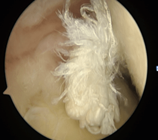
Figure 2: Torn LHB in shoulder joint viewed with arthroscope
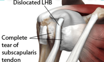
Figure 3: Dislocated LHB with concomitant rotator cuff tear
What are Common Treatments for Biceps Tendon Disorders?
For the treatment of long head biceps tendinitis and tendinosis, an initial course of NSAID’s, avoidance of offending activities such as throwing and heavy lifting along with physical therapy may be successful in alleviating pain. An intra-articular corticosteroid injection may help alleviate symptoms temporarily or for the long term. In cases of instability of the LHB and failed long term symptom relief surgical options may be reasonable. Tenotomy, is the release of the LHB from its attachment site in the shoulder and can result in successful pain relief. Tenodesis is a surgical procedure transferring the LHB from its attachment site in the shoulder joint and re-attachment of the tendon to the upper arm bone (humerus) (Fig 4). Both procedures have been shown in scientific controlled trials to result in similar functional outcomes. The advantage of the tenotomy is that patients do not require protected rehab following LHB release in contrast to a tenodesis. The disadvantages of tenotomy are the possibility of a cosmetic “popeye” muscle deformity and some muscle cramping with vigorous repetitive resistance activities. In patients with proven symptomatic instability of the LHB by MRI imaging, tenotomy or tenodesis is the treatment of choice rather than prolonged non-op measures given the associated risk of damage to the rotator cuff (Fig 3). In the setting of an acute rupture of the LHB at the shoulder, treatment is typically non-operative given the similar outcomes of tenotomy and tenodesis. In contrast to LHB ruptures at the shoulder , distal biceps ruptures at the elbow have better outcomes when surgically repaired early. Delayed repairs greater than three weeks may require tendon grafting to restore the re-attachment of the biceps to the elbow resulting in outcomes inferior to early direct repair (Fig 5).
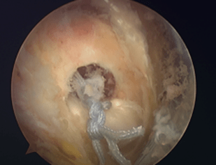
Figure 4 : Arthroscopic view of LHB tenodesis.
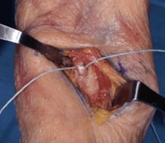
Figure 5: Distal biceps undergoing operative repair to bone.
